Saint Francis Hospital Continues to Lead the Way in Interventional Pulmonology
August 23, 2023Categories: Organizational Updates
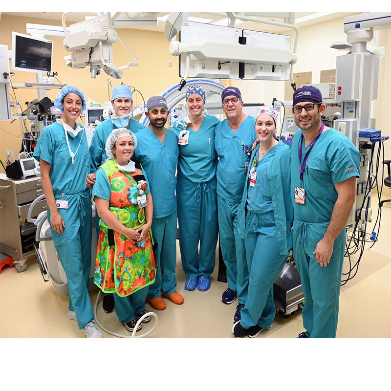
Saint Francis Hospital is once again leading the way in providing state-of-the-art care, becoming the first hospital in Connecticut to combine Cios Spin with the Ion endoluminal system for robotic bronchoscopy. Anil Magee, M.D., Director of Interventional Pulmonology, and the interventional pulmonology team are thrilled to be able to provide more precise procedures for patients with lung nodules, which ultimately leads to faster diagnosis and subsequent treatment.
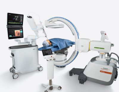
Cios Spin is a state-of-the-art mobile 3D imaging system which allows Dr. Magge to pinpoint the exact location of a suspicious lung nodule. Previously, a CT scan of the chest was taken prior to an Ion procedure to map a pathway to the nodule. By integrating Cios Spin with Ion, Dr. Magge is now able to navigate within the lung with even greater precision, thanks to the real-time, CT-like images, which allow readjustments if needed.
“Performing lung nodule biopsies through the airway is not only safer for the patient, but this system also allows us to determine a stage of cancer as well as ruling in or out the possibility of cancer in the lymph nodes,” said Dr. Magge. “Combining Ion and Cios Spin is helping us provide life-changing care to our patients. Detecting and treating cancerous nodules earlier greatly improves patient outcomes. We are excited to be the first in the state to provide these minimally invasive diagnostics in a single procedure.”
Dr. Magge and the interventional pulmonology team were the first in Connecticut to use the Ion system last October and have since completed over 250 procedures for patients in need of lung biopsies in difficult to reach areas of the lung. The Ion system features an ultra-thin, ultra-maneuverable catheter that allows navigation far into the lung, addressing a challenging aspect of lung biopsy. Along with unparalleled stability, this enables the precision needed for biopsy compared to other technologies.
During bronchoscopy with Ion, Dr. Magge uses a console to navigate to the target within the lung along a pre-planned pathway. The catheter can move 180° in any direction to pass through small, difficult to navigate airways to reach the lung nodule of concern. Ion’s peripheral vision probe provides direct vision during navigation. Once at the desired location, the catheter locks into place and biopsy tools are then passed through the catheter to take a sample of the nodule.